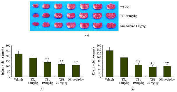
Figure 1.
(a) Representative coronal brain sections stained with TTC after 2 hours of MCAO and 24 hours of reperfusion showing infarction. Dark-colored region in the TTC stained sections indicated nonischemic portion of brain and pale-colored region indicated ischemic portion of brain. Theaflavin and nimodipine-treatment reduced infarct volume. (b) Volume of infarction after 2 hours of MCAO and 24 hours of reperfusion in vehicle, theaflavin (5, 10, and 20 mg/kg) and nimodipine (1 mg/kg)-treated rats. (c) Volume of edema after 2 hours of MCAO and 24 hours of reperfusion in vehicle, theaflavin (5, 10, and 20 mg/kg) and nimodipine-treated (1 mg/kg) rats, **P < .01 as compared to the vehicle-treated group.

