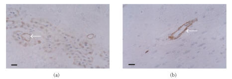
Figure 2.
Immunohistochemical staining of ICAM-1 in brain tissues of (a) vehicle-treated rats and (b) theaflavin-treated rats (20 mg/kg), SP×400. ICAM-1 protein is mainly expressed on the microvascular endothelial cells. ICAM-1 expression decreases dramatically in theaflavin-treated groups. Scale bar = 10 μm.

