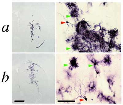
Figure 6.
Expression of β-galactosidase seen 6 months after injection of RAd35 (row a) or AdGS46 (row b). Animals were injected with 1.3 × 10⁷ infectious units of vector and were left to survive for 6 months without re-exposure to adenovirus. Brains were subsequently analyzed by immunohistochemistry for β-galactosidase immunoreactivity. Red arrowheads delineate transduced cells with neuronal morphology and green arrowheads delineate astroglial-like cells (the predominant cell-type transduced). [Bar = 1 mm (low magnification images) and 100 μm (high magnification images).]

