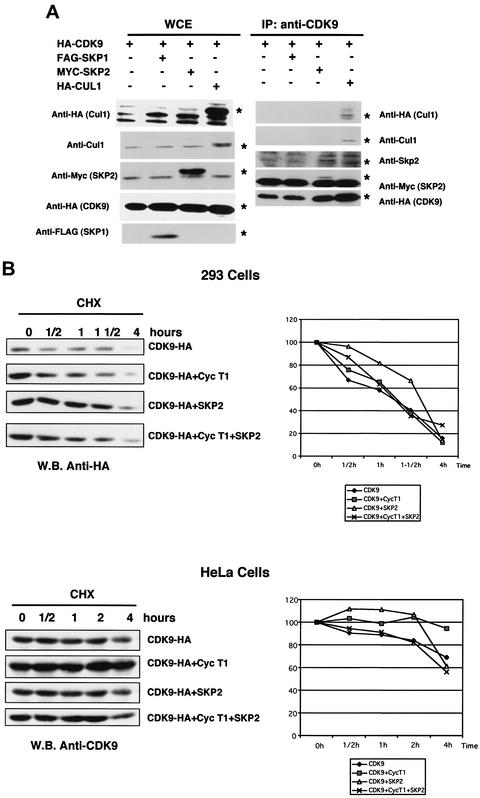
FIG. 5.
CDK9 interacts with SKP2 and Cul-1 in transfected 293 cells. (A) Cells from the 293 cell line were transfected with pCMV-CDK9-HA (HA-CDK9), pCMV-Flag-SKP1 (FAG-SKP1), pCMV-myc-SKP2 (MYC-SKP2), and pCMV-HA-Cul1 (HA-Cul1) in the combinations indicated. Five micrograms of each plasmid was transfected together with 0.5 μg of a luciferase reporter plasmid by the calcium phosphate method. The cells were collected 48 h following transfection, and whole-cell extracts were immunoprecipitated with anti-CDK9 antibodies. Whole-cell extracts and immunoprecipitates were resolved by SDS-PAGE followed by Western blot analysis. The left panels show the expression levels of the transfected proteins, determined by using specific antibodies. The right panels show coimmunoprecipitation of Cul-1 and SKP2 with CDK9. SKP1 could not be detected in this experiment (see text for details). Asterisks indicate the migration of specific bands. WCE, whole-cell extracts; IP, immunoprecipitates. (B) Ectopic expression of SKP2 and cyclin T1 alone or in combination does not affect CDK9 t1/2. CDK9-, SKP2-, and cyclin T1-expressing plasmids were transfected into 293 cells alone or in combination, as indicated in the upper panels. In parallel experiments, HeLa cells were transduced with Ads expressing CDK9, SKP2, and cyclin T1 alone or in combination (lower panels). Twenty-four hours posttransfection and posttransduction, the cells were divided onto five identical plates. Twenty-four hours later, the cells were incubated with CHX (40 μg/ml) for the indicated periods of time. The expression levels of CDK9 at each time point were analyzed by Western blotting. In each case, Western blots from a representative experiment as well as quantification of the results of three independent experiments are shown. Expression levels are represented as percentages of the expression values at time zero. W.B., Western blot.

