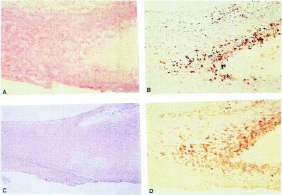
Figure 7.
Serial sections of carotid arteries obtained by endarterectomy were processed as described in Methods. Rabbit polyclonal antibodies produced against an apoB48R-specific domain (amino acids 223–710) GST-fusion protein were used to determine apoB48R expression and location. (A) Hematoxylin-stained section of an advanced lesion; a lipid core surrounded by foam cells is seen to the right and center (×100). (B) A macrophage-specific monoclonal antibody (HAM56) identifies the macrophage-derived foam cells around the lipid core (×100). (C) Preimmune IgG from the rabbit used to generate the anti-apoB48R IgGs did not bind to lesion cells (negative control) (×80). (D) Anti-apoB48R IgGs specifically bind to lesion macrophages and foam cells (×100), indicating these cells express the apoB48R protein.

