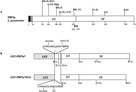
FIG. 1.
Schematic diagrams of S. pneumoniae PBP2a. (A) Topology of the native PBP2a protein. The solid and hatched boxes indicate the N-terminal cytoplasmic region and the membrane anchor, respectively. GT and TP domains are represented together with their conserved motifs; active site serine 410 is indicated by an asterisk. x represents any amino acid. The gray box and the arrows indicate the location of the identified GT and TP junction (5). (B) Schematic diagrams of the PBP2a-derived constructs. The peptide at the GST-GT junction of the PBP2a* construct includes sequences specific for thrombin, Tev protease, and factor Xa (italic characters) and N-terminal amino acids of GT (bold characters).

