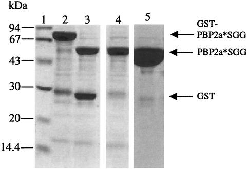
FIG. 2.
Purification of PBP2a*SGG. Proteins were separated by sodium dodecyl sulfate-12.5% PAGE and stained with Coomassie blue. Numbers at the left indicate sizes of standard molecular mass markers. Lanes: 1, molecular mass markers; 2, GST-PBP2a*SGG fusion following purification in a glutathione-Sepharose column; 3, fusion protein cleavage by Tev protease; 4, flowthrough from glutathione-Sepharose (10 μg of protein loaded on the gel); 5, elution from Resource Q column (30 μg of protein loaded on the gel).

