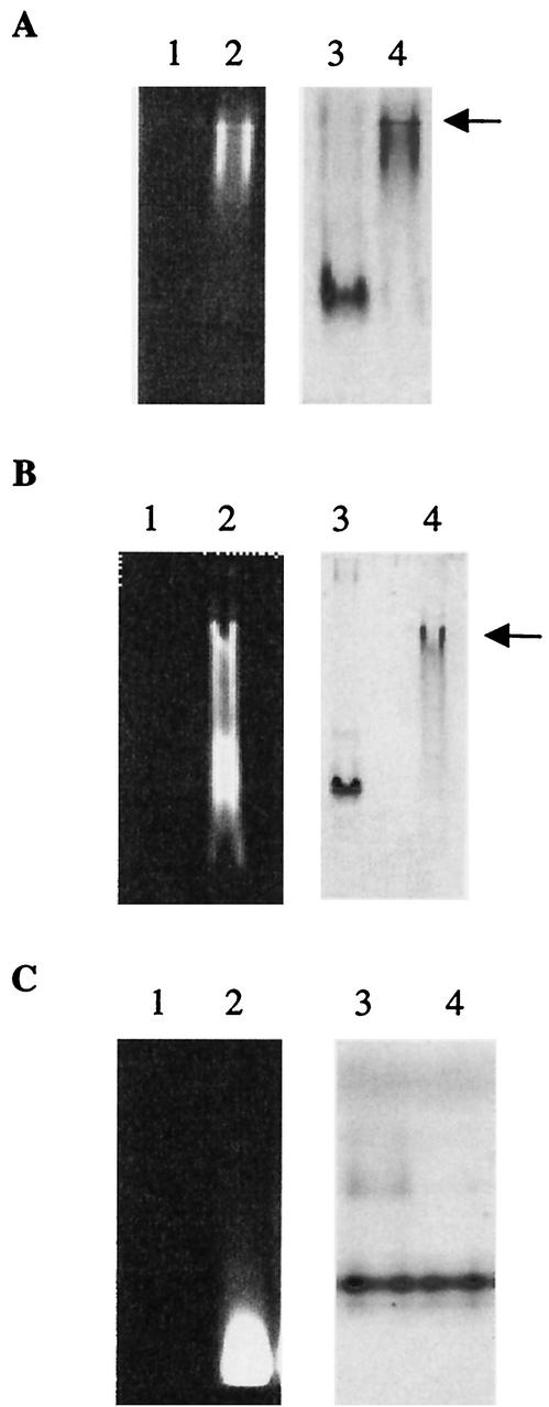
FIG. 3.
Lipid II binding to PBP2a moieties. The binding was performed at room temperature for 15 min before loading onto native PAGE. Arrows indicate protein/lipid II complex. (A) Lanes: 1 and 3, PBP2a* (2 μM); 2 and 4, PBP2a* (2 μM) and lipid II (140 μM). The same gel was observed under UV light (lanes 1 and 2) before staining with Coomassie blue (lanes 3 and 4). (B) Lanes: 1 and 3, PBP2a*SGG (2.8 μM); 2 and 4, PBP2a*SGG (2.8 μM) and lipid II (140 μM). The same gel was observed under UV light (lanes 1 and 2) before staining with Coomassie blue (lanes 3 and 4). (C) Lanes: 1 and 3, TP2a (5.5 μM); 2 and 4, TP2a (5.5 μM) and lipid II (140 μM). The same gel was observed under UV light (lanes 1 and 2) before staining with Coomassie blue (lanes 3 and 4).

