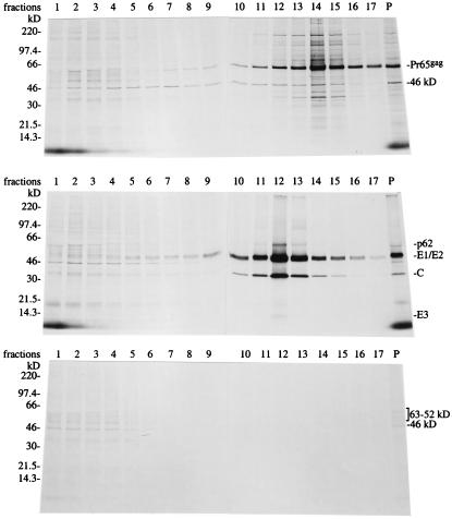
Figure 1.
Purification of Gag particles. (Top) Cells (4 × 10⁶) were infected with SFV-C/Pr65gag vectors and labeled with [35S]methionine both before and after infection. Released particles in medium were collected between 5.5 and 6.0 h after infection and analyzed by sedimentation on a 5–20% iodixanol gradient. Particles were recovered from each fraction by pelleting and analyzed by SDS/PAGE. Autoradiographies of the gels are shown. Major proteins are indicated. P, pellet in gradient. (Middle) Cells were infected with SFV. Labeling of cells and particle analysis were as described above. Note that the iodixanol gradient was in this case 5–30%. (Bottom) Cells were infected with SFV-C/NP vectors. Labeling of cells and analysis of medium were as described for Top.

