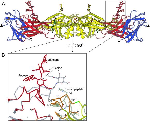
Fig. 2.
The glycan at residue 153 in dengue 2 virus E protein. (A) The E protein dimer, viewed perpendicular to the dyad axis (and the view in Fig. 1 A). Both glycans are approximately perpendicular to the viral surface. Domain I and the attached glycan are shown in red, domain II and the attached glycan are shown in yellow, and domain III is in blue. Disulfide bridges are shown in orange. The molecule of β-OG bound in the hydrophobic pocket underneath the kl hairpin is in green. A putative receptor-binding loop in domain III (residues 382–385) is marked with a triangle. (B) Enlargement of the area surrounding the glycan at residue 153 in domain I, with the structure of TBE envelope protein superimposed (gray) onto domain I of dengue virus E protein. The fusion peptide is highlighted in orange. The disulfide bridge between residues 92 and 105 is shown in green.

