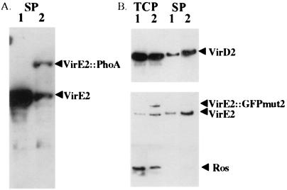
Figure 2.
Western blots of A. tumefaciens containing VirE2 fusions with PhoA and GFPmut2. (A) Supernatant fraction from PhoA constructs detected with VirE2 antibodies. A348 (lane 1) was grown under inducing conditions, and A136 (phoA−) (ptac-virE1 + E2∷phoA/pUFR047) (lane 2) was grown under noninducing conditions. (B) Cells were grown at 28°C under inducing conditions, separated into the supernatant and total cell fractions (noted on the top), and detected with protein-specific antibodies (noted on the right). Lane 1, A348 (pUFR047); and lane 2, A348 (ptac-virE1 + E2∷gfpmut2).

