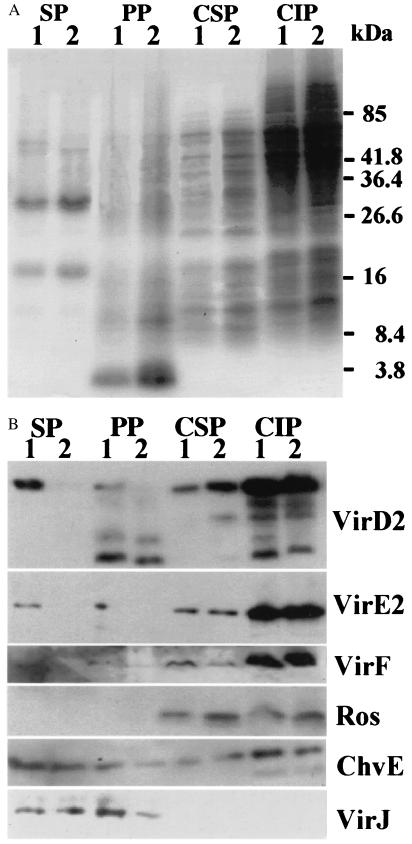
Figure 4.
Coomassie Brilliant Blue staining and Western blotting of protein gels from different fractions of A. tumefaciens cells containing ptac-osa. (A) Cells were grown under inducing conditions and then collected, and separated into supernatant, periplasmic, cytoplasmic-soluble, and cytoplasmic-insoluble fractions (noted on the top). The fractions were run on SDS/PAGE gels, and stained with Coomassie Brilliant Blue. Lane 1, A348 (pUFR047); and lane 2, A348 (ptac-osa/pUFR047). (B) Western blots of the proteins from different fractions (noted on the top) detected with antibodies (noted on the right). Lane 1, A348 (pUFR047); and lane 2, A348 (ptac-osa/pUFR047). Periplasmic proteins, PP; cytoplasmic-soluble proteins, CSP; and cytoplasmic-insoluble proteins, CIP.

