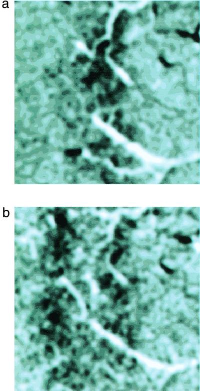
Figure 3.
The optical image of a flickering bar. (a) Image of the area V-1 cortical intrinsic signal generated by a flickering bar 50 ms on and 100 ms off, that was 0.13° wide with an orientation of 132°. The patch is 1 cm2 and was approximately 10–12° below and to the left of the foveal presentation and subtended about 4° of visual angle (as measured with microelectrode penetrations at each edge of the image), at the anterior-medial border of the operculum. The vertical meridian is parallel to the lower edge of this image; the fovea is to the right. (b) Image of the intrinsic signal generated by a flickering bar 0.64° wide in the same piece of cortex (the center of the bar was also shifted here approximately 0.29° away from the fovea). Notice that the widened bar has shifted in position and split into two edges.

