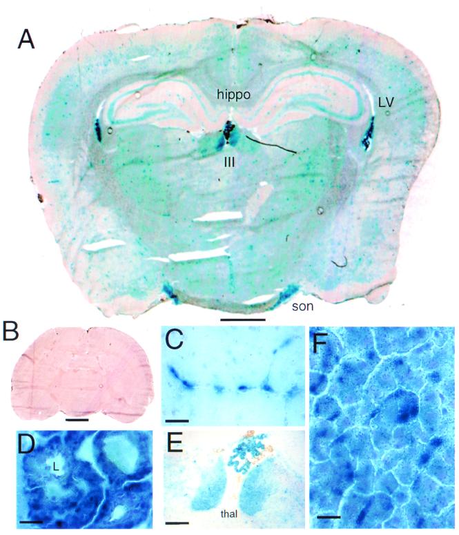
Figure 5.
β-galactosidase histochemistry in brain (A–E) and liver (F) at 48 h after i.v. injection of the β-galactosidase gene packaged inside the OX26 pegylated immunoliposome (B–F). The control brain from rats receiving no gene administration is shown in B. Magnification bars = 1.5 mm (A), 2.2 mm (B), 57 μm (C), 23 μm (D), 230 μm (E), and 15 μm (F). A and B were not counterstained. The lateral ventricles (LV), third ventricle (III), left or right hippocampus (hippo), and hypothalamic supraoptic nuclei (son) are labeled in A. C shows punctate gene expression in intraparenchymal capillaries and may represent gene expression in either endothelium or microvascular pericytes. (D) Gene expression in the epithelium of the choroid plexus. The lumen (L) of a capillary of the choroid plexus is shown and demonstrates the absence of β-galactosidase enzyme activity in the plasma compartment. (E) The thalamic (thal) nuclei below the choroid plexus of the third ventricle, which is also visible in A. (F) Abundant gene expression in hepatocytes; this high magnification shows a speckled pattern, suggesting localization of the β-galactosidase enzyme within the liver cell endoplasmic reticulum.

