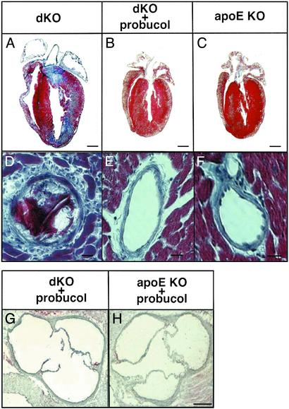
Fig. 3.
Histologic analysis of hearts. (A–F) Representative longitudinal sections of hearts from 6-week-old untreated dKO (A and D), probucol-treated dKO-P (B and E), and untreated apoE single KO (C and F) mice stained with Masson's trichrome (healthy myocardium, red; fibrotic infarcted tissue, blue). (G and H) Oil red O-stained sections through the aortic sinuses of probucol-treated dKO (G) and apoE KO (H) mice. (Scale bars: A–C, 1 mm; D–F,10 μm; G and H, 100 μm.)

