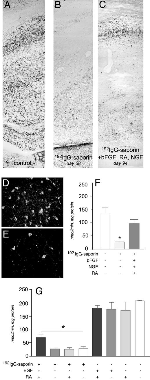
Fig. 3.
Cholinergic denervation in the hippocampus of 192IgG-lesioned animals and effects of combined treatments. Histochemical visualization of acetylcholinesterase in the hippocampal cortex of control rats (A) and 68 days after injection (B) is shown. The immunolesion induces an almost complete disappearance of esterase-reactive fibers in the hippocampus by day 68, and the deafferentation does not further worsen at 85 days (data not shown). Degeneration of cholinergic neurons in the basal forebrain is also shown (D, control, sham-operated; E, lesioned). (C) Esterase-reactive fibers were more abundant in lesioned animals after bFGF, retinoic acid (RA), and NGF administration (94 days). ChAT activity also drops in the hippocampus of lesioned animals, and combined treatment with mitogens, retinoic acid, and NGF recovers this deficit (F). In addition, the mitogens combined with retinoic acid partially recover the ChAT deficit (G), whereas single treatments do not modify ChAT activity in either lesioned or control animals. Statistical analysis was carried out by using ANOVA and Dunnett's test (P < 0.05).

