Figure 1.
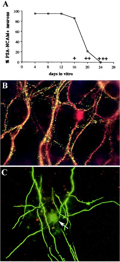
Down-regulation of neuronal PSA-NCAM is concomitant with the onset of myelination. Cultures derived from embryonic (E15) mouse hemispheres were maintained for various periods of time before being double-labeled with anti-PSA-NCAM and either TuJ1 to label neurons (B) or anti-MBP mAb to label myelinated internodes and oligodendrocytes (C). (A) The percentage of PSA-NCAM-expressing neurons (TuJ1-positive cells) is plotted as a function of time in culture. The intensity of myelination in sister cultures was evaluated by counting the number of myelinated MBP positive internodes and is indicated by + (onset), ++ (moderate), and +++ (maximum). (B) Immunostaining of TuJ1-positive neurons (red) with anti-PSA-NCAM mAb (green) at 16 DIV. Most of the neurites appear in yellow because of the superposition of the two labeling. (C) A field of myelinated internodes linked to a single oligodendrocyte at 24 DIV. Only rare PSA-NCAM dots (red) are visible, contrasting with the intense labeling of MBP-positive myelinated internodes (green). Arrow points to the myelinating oligodendrocyte cell body, which is not in the same plane of focus and is moderately stained with the anti-MBP mAb, as after the onset of myelination, most of the MBP migrates out the oligodendrocyte cell body. (B and C, magnification ×320.)

