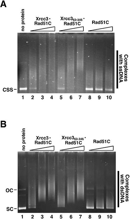
Figure 3.
DNA-binding activity of Xrcc363–346–Rad51C. (A) ssDNA binding. Single-stranded pGsat4 DNA (40 µM) was mixed with the indicated amounts of Xrcc3–Rad51C, Xrcc363–346–Rad51C or Rad51C in 10 µl of the standard reaction buffer. The reaction mixtures were incubated at 37°C for 10 min and were analyzed by 0.8% agarose gel electrophoresis. The bands were visualized by ethidium bromide staining. (B) dsDNA binding. Superhelical pGsat4 DNA (15 µM; 3216 bp) was mixed with the indicated amounts of Xrcc3–Rad51C, Xrcc363–346–Rad51C and Rad51C in 10 µl of the standard reaction buffer. The reaction mixtures were incubated at 37°C for 10 min and the samples were analyzed by 0.8% agarose gel electrophoresis. The bands were visualized by ethidium bromide staining. Lane 1 indicates the experiment without protein. Lanes 2–4 indicate the experiments with Xrcc3–Rad51C. Lanes 5–7 indicate the experiments with Xrcc363–346–Rad51C. Lanes 8–10 indicate the experiments with Rad51C. Protein concentrations were 0.35 (lanes 2, 5 and 8), 0.7 (lanes 3, 6 and 9) and 1.1 µM (lanes 4, 7 and 10).

