Figure 3.
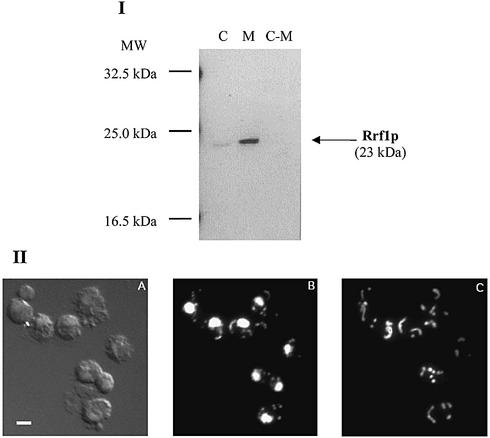
(I) Rrf1p is localized in the fraction containing mitochondria. Western blot analysis of crude yeast extracts (designated column C), isolated mitochondria (column M) and post-mitochondrial supernatant fraction (C-M) obtained from wild-type strain DS413. Proteins (40 µg) of each preparation were analyzed by western blotting using antibody against His6-partial Rrf1p. (II) Cytological localization of Rrf1p to mitochondria. A representative microscopic field of Δrrf1 rho+ haploid cells (strain ET2) harboring a high copy number plasmid carrying RRF1 (pRRF1W2) is shown. (A) Differential interference contrast view. (B) DAPI staining showing the locations of nuclear and mitochondrial DNA. (C) Indirect immunofluorescence using antibodies to His6-partial Rrf1p and FITC- conjugated secondary antibody. Bar represents 1 µm.

