
Figure 4.
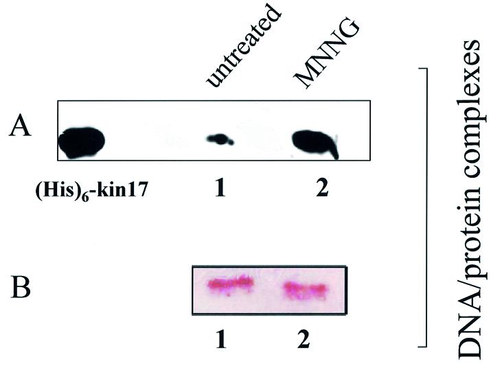
kin17 protein detection in purified protein–chromatin DNA and protein–protein complexes after in vivo cross-linking of living S phase HeLa cells with formaldehyde. After in vivo cross-linking with formaldehyde (1%, 4 min), the nuclei were isolated, lysed and both protein–chromatin DNA and protein–protein complexes were purified by equilibrium centrifugation in two consecutive cesium chloride gradients as described in Materials and Methods. After reversion of the cross-links by boiling, immunoblot analysis was performed on DNA–protein complexes obtained from untreated (A and B, lanes 1) and MNNG-treated (A and B, lanes 2) HeLa cells. (A) Treatment of cells for 3 h with 100 µM MNNG immediately after release from a double thymidine block. Detection of kin17 protein. (B) Detection of histone H1. (His)6-kin17, positive control [recombinant human (His)6-kin17 protein].