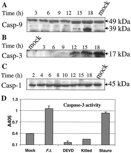
FIG. 2.
(A to C) Kinetic analysis of caspase activation from proteolysis in J774A.1 macrophages after Francisella infection. Cells were infected and collected, and cell extracts were prepared at the indicated time points (in hours; indicated over the blots) and analyzed by Western blot analysis. (A) Involvement of caspase 9 (Casp 9) in the F. tularensis-induced apoptotic pathway of J774A.1 macrophages. The 39-kDa fragment represents the cleaved product. (B) Cleavage of procaspase 3 into active caspase 3 in F. tularensis-infected J774A.1 macrophages. Cell lysates were analyzed for cleavage of procaspase 3 using immunoblots with anti-caspase 3 antibodies. (C) No detectable cleavage of caspase 1 in infected J774A.1 macrophages. (D) Caspase 3 activity in F. tularensis-infected cells. Positive-control cells were treated with 1 μg of staurosporine (Stauro) ml−1 for 12 h. The other samples were processed at 18 h. The cells were treated as follows: not infected (Mock), infected with F. tularensis (F.t.), infected plus treated with Ac-DEVD-DEVD (fluoromethyl ketone [fmk]), and treated with killed F. tularensis (Killed). Values are the means ± standard deviations (error bars) from three samples (error bars of mock-infected cells and cells treated with killed F. tularensis not visible).

