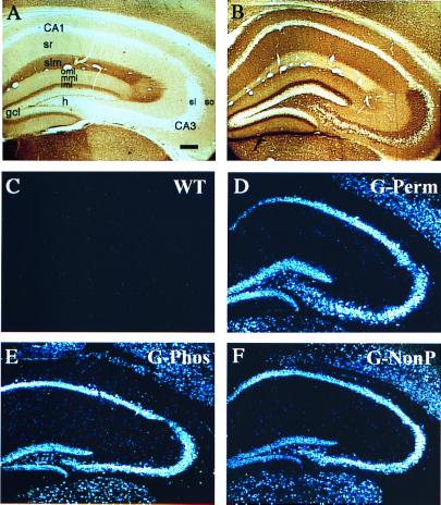
Figure 3.
(A and B) Immunohistochemical staining with 7B10 Ab, which recognizes both the endogenous and the transgenic GAP-43 protein in a WT (A) and G-Perm (B) mouse. Note that, in dentate gyrus, increased staining is observed in the perforant path target zone (oml and mml, outer and middle molecular layers, respectively), the mossy cell target zone (iml, inner molecular layer), and the mossy fibers (stratum lucidum). (C–F) Similar GAP-43 overexpression among the three transgenic mouse lines in the major cell fields in hippocampus. Darkfield photomicrographs of in situ hybridization with riboprobe that recognizes transgenic chick GAP-43 mRNA but not endogenous mouse GAP-43 mRNA. Note similar levels of expression in the three transgenic lines (D–F) in the major subfields of the hippocampus and absence of transgene in WT mice (C). Abbreviations: h, hilus; so, stratum oriens; slm, stratum lacunosum moleculare; sr, stratum radiatum; gcl, granule cell layer. Bar = 150 μm.

