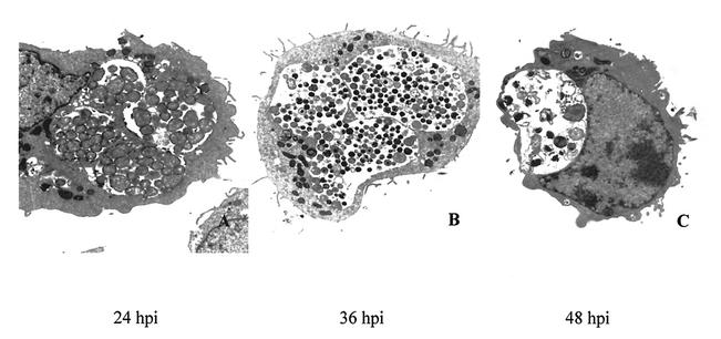
FIG. 1.
Ultrastructural analysis of C. suis S-45 growth in infected BGMK cells. Transmission electron photomicrographs of BGMK cells infected with C. suis S-45 at 24 (A), 36 (B), and 48 (C) hpi. (A) RBs predominated in multiple inclusions at 24 hpi. (B) By 36 hpi, inclusions contained a mixture of RBs and mature EBs. (C) Essentially empty or partially empty inclusions were visualized at 48 hpi, and the remaining EBs and RBs in these inclusions appeared aberrant or damaged. Magnification, ×8,000.

