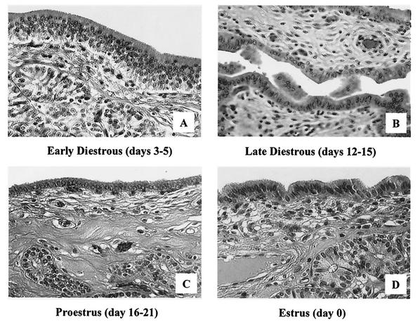
FIG. 2.
Histological analysis by light microscopy of hematoxylin-and-eosin-stained sections of swine endometrial tissue during the estrus cycle. (A) Tissue at early diestrus (days 3 to 5) shows a high columnar luminal epithelium, a large number of glandular epithelial cell clusters that are close to the luminal epithelium, and increased epithelial mitotic activity. (B) Tissue at late diestrus (days 12 to 15) reflects shedding of many luminal epithelium cells and a transition from high columnar epithelia to the low columnar form. (C) In proestrus (days 16 to 21), single cubical epithelia dominate the luminal surface and glandular clusters are located deep in the stromal layer. (D) Tissue at estrus (day 0) shows a high columnar luminal epithelium and a large number of glandular epithelial cell clusters that are close to the luminal epithelium.

