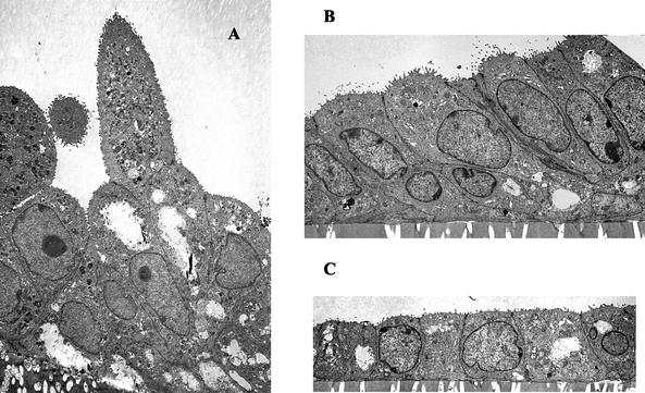
FIG. 3.
Ultrastructural analysis of polarized primary swine glandular epithelial cells. (A) Tissue at early diestrus (days 3 to 5) illustrates tall columnar epithelial cells with high miotic activity and the formation of characteristic gland-like (organoid) structures. (B) Tissue at late diestrus (days 12 to 15) reflects high columnar epithelia. (C) In tissue at proestrus (days 16 to 21), the monolayer is representative of single cuboidal epithelial cells. Magnification, ×2,900.

