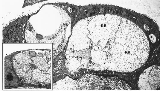
FIG. 7.
Ultrastructural analysis of C. suis S-45 in estrogen-dominant primary swine uterine glandular epithelial cells. Shown are transmission electron photomicrographs of primary glandular epithelial cells infected with C. suis S-45 at 40 hpi; abnormally enlarged RBs predominated in inclusions formed in these cells. Magnification, ×2,100. (Inset) Portion of an inclusion containing a mixture of enlarged as well as normal RBs and some mature EBs. Magnification, ×4,200.

