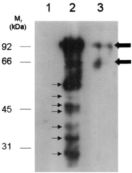
FIG. 4.
Protein blot analysis of mycoplasma cell surface S-plasminogen binding proteins from strain PG50. Cells were incubated either in the absence of S-plasminogen (lane 1), in the presence of S-plasminogen alone (lane 2), or in the presence of S-plasminogen and 1 mM TA (lane 3) prior to UV exposure. After UV exposure (which is required to transfer biotin to plasminogen binding proteins), whole-cell lysates were prepared and fractionated by reducing SDS-12% PAGE (approximately 15 μg/lane), and the proteins were transferred to PVDF membranes. The blot shows biotin-labeled lysine-dependent plasminogen binding proteins (thin arrows) detected with neutravidin-HRP followed by chemiluminescence. Thick arrows indicate traces of S-plasminogen detected with neutravidin-HRP. Molecular masses of molecular mass markers are as indicated.

