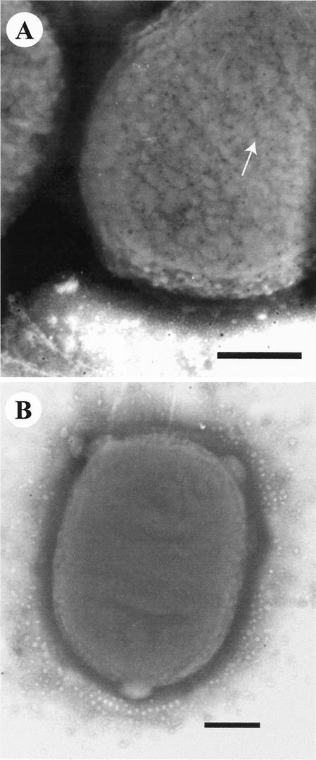
FIG. 7.
Transmission electron micrographs showing the uniform distribution of immunogold-labeled Paa protein (arrow) over the bacterial surface of the complemented strain M155c (A) following overnight growth at 37°C in TSB. When anti-Paa serum was adsorbed against the Paa protein, only a few gold beads were observed for strain M155c, mostly in the background (B). Bars = 300 nm.

