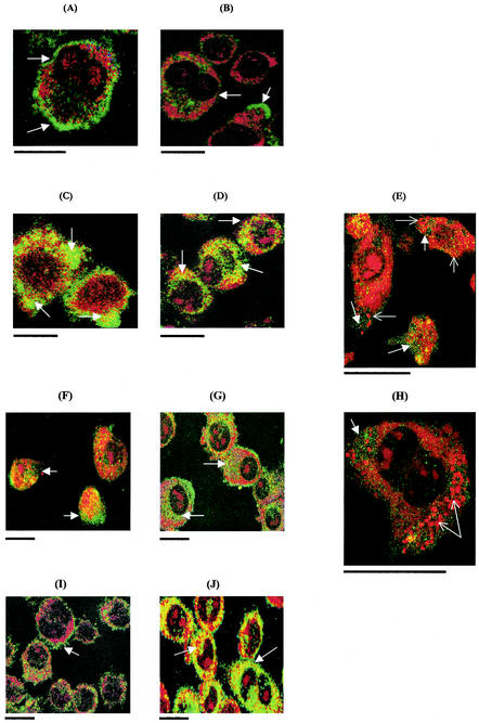
FIG.3.
Confocal laser scanning microscope images of gp91phox activity in J774.2 macrophages infected with salmonellae and cocultured with or without IFN-γ (1,000 U/ml). (A) Uninfected control, 2 h in culture; (B) uninfected control, 12 h in culture; (C) 14028 phoP mutant, 12 h postinfection, without IFN-γ; (D) 14028 phoP, 12 h postinfection, with IFN-γ; (E) high-power image of 14028 phoP mutant-infected cell, 12 h postinfection, without IFN-γ (image taken 4 μm below cell surface); (F) wild-type 14028, 12 h postinfection, without IFN-γ; (G) wild-type 14028, 12 h postinfection, with IFN-γ; (H) high-power image of wild-type 14028, 12 h postinfection, without IFN-γ (4 μm below cell surface); (I) IFN-γ only, 2 h in culture; (J) IFN-γ only, 12 h in culture. Yellow-green shows gp91phox activity. Open arrow shows gp91phox localization on cell membrane; solid arrow shows gp91phox cytoplasmic localization. Solid arrows in E and H show bacteria in cells. Scale bars, 10 μm.

