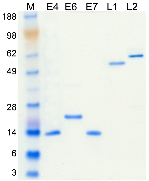
Figure 1.
Analysis of purified proteins by SDS-PAGE. The purified HPV16 proteins E4, E6, E7, L1 and L2 were run on a polyacrilamide gel electrophoresis and stained by Coomassie blue. Each protein is indicated on the top of the corresponding lane. The weight of the molecular mass markers (lane M) is indicated on the left of the figure.

