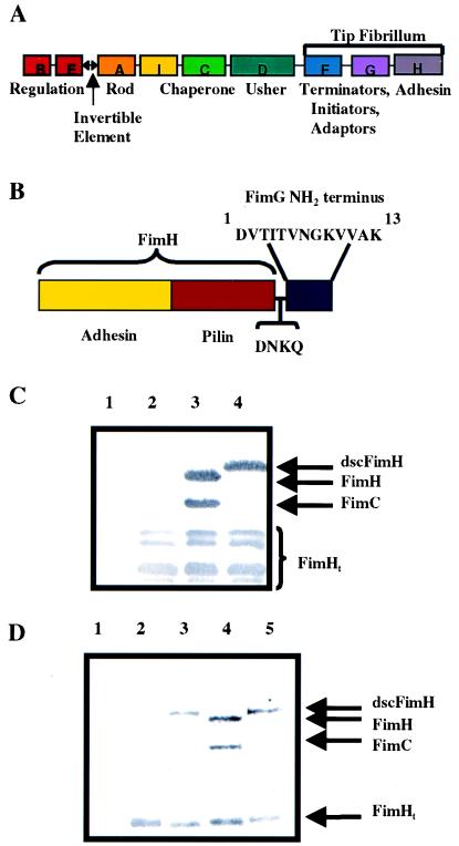
Figure 1.
Donor strand complementation of FimH in cis. Shown are schematic diagrams of the type 1 gene cluster (A) and dscFimH (B). Immunoblots developed with anti-FimCH antiserum of periplasmic extracts (C) after no expression of FimH (lane 1), FimH alone (lane 2), FimH + FimC (lane 3), or dscFimH (lane 4). A proportion of FimH truncation occurred under all conditions and was labeled FimHt. (D) Elution of FimH or dscFimH from mannose-Sepharose after incubation with periplasm containing FimC (lane 1), FimH alone (lane 2), dscFimH (lane 3), FimH + FimC (lane 4), or dscFimH + FimC (lane 5). The elutions were run on a SDS/PAGE gel followed by Western blotting using anti-FimCH antibodies.

