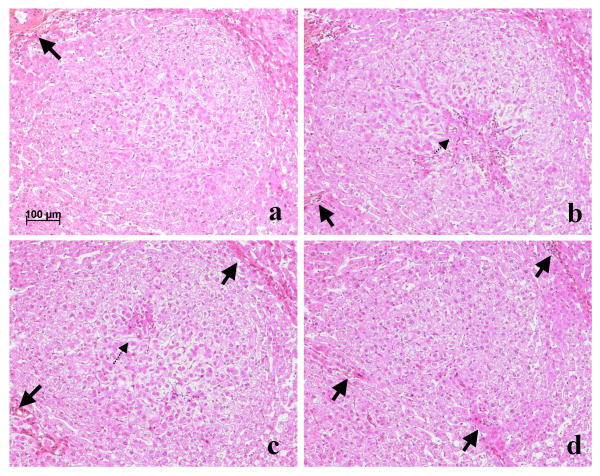
Figure 4.
(a, b, c, d), Patient 4, nodule b: type 4 lesion, serial sections. The hyperplastic focus is surrounded by atrophic hepatocytes and dilated sinusoids in which course arteries or arterioles (thick arrows). According to the plane of section, regions of regenerating hepatocytes, associated with ductular reaction (not visible at this magnification), and feeding arteries (dotted arrow) are more or less obvious. All, H&E.

