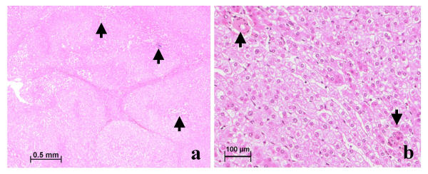
Figure 5.
Patient 4, nodule b (same nodule as Fig 4, another region): type 3–4 lesion. (a) Fibrotic bands without ductular reaction encircle hyperplastic foci; arterioles in the center or at the periphery (arrow) are cuffed by a ductular reaction (not visible). Sinusoids along fibrotic bands are slightly dilated and a large region of sinusoidal dilatation can be seen on the left. H&E. (b) Between 2 hyperplastic foci is a zone of mild hepatocytic atrophy and sinusoidal dilatation in which arteries (arrow) course. H&E.

