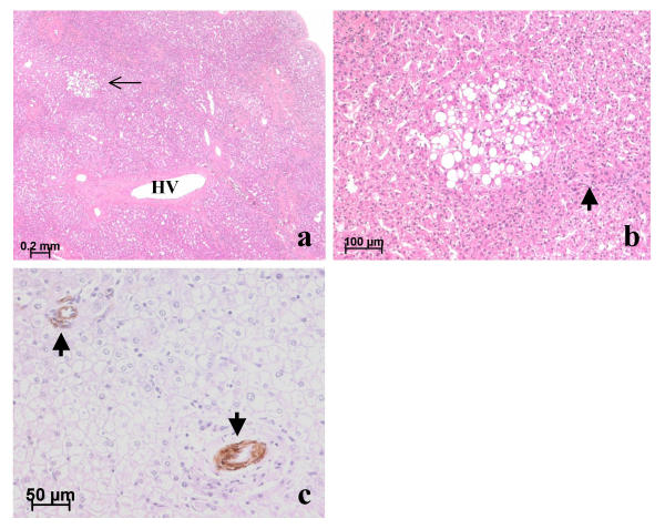
Figure 7.
Non-nodular liver: (a, b) Patient 7; (c) Patient 3. (a) Near a steatotic nodule (not shown) lies a region of abnormal parenchyma. Note approximated portal tracts with thick-walled arteries and bile ducts (not visible at this magnification) but lacking portal veins, a large hepatic vein (HV) with a very thick wall, and a steatotic focus (thin arrow). (b) At a higher magnification, this steatotic focus near an artery (thick arrow) is partly bordered by slightly dilated sinusoids. H&E. (c) On the left, the portal tract contains an artery (arrow) but no visible bile duct; on the right an artery not partnered with a bile duct. Anti-α-SMA immunostaining.

