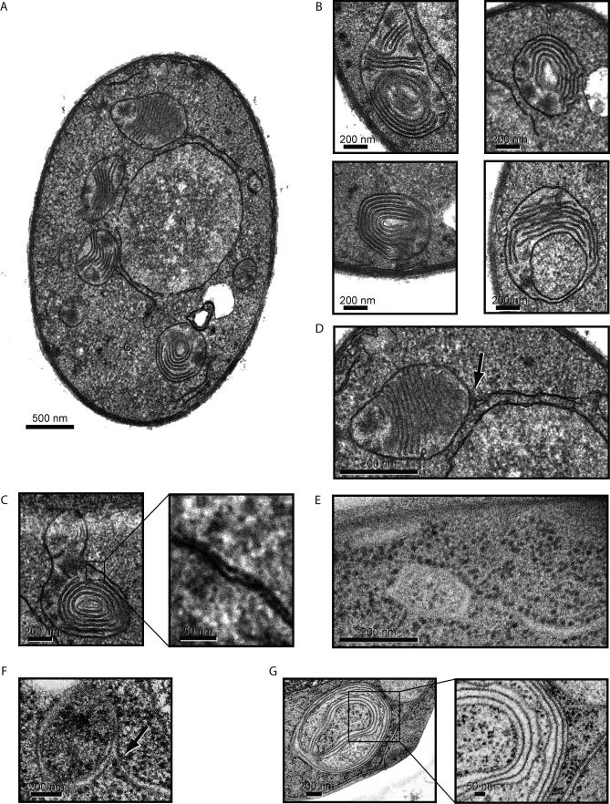
Figure 3. Characterization of ER-Containing Autophagosomes (ERAs) during the UPR.
(A) Images of representative DTT-treated wild-type cells that contain ERAs. Nuclei and cytoplasm are indicated as N and C, respectively.
(B) Enlargement of representative images of ERAs from different cells. The bottom right image is likely to show a section through a cup-shaped ERA. Note that there are no connections between the stacked cisternae and the envelope.
(C) High magnification of the ERA double membrane envelope.
(D) Some ERAs are found attached to or are in close proximity to ER tubules/sheets (indicated by the arrow). Note that the section in (A) includes two such junctions.
(E) High-pressure freezing/freeze substitution image of an ERA linked to an ER tubule/sheet. The osmium/lead staining used in this technique visualizes ribosomes and demonstrates that the outer ERA envelope membrane, but not the stacked internal cisternae, are tightly studded with ribosomes, indicating that they originate from ER membranes.
(F) High-pressure freezing/freeze substitution image of an ER-ERA junction using an improved protocol to visualize membranes.
(G) Using the same technique as in (F), we visualized the internal membrane content of an ERA. Note that both portions of the internal membranes and of the sequestering double membrane envelope contain bound ribosomes, and hence are likely derived from the ER.

