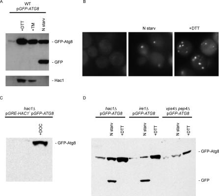
Figure 6. UPR-Induction of the Autophagy Marker GFP-Atg8.
(A) Wild-type cells transformed with a plasmid containing GFP-Atg8 were grown for 4 h in synthetic media with no drug, with UPR-inducing conditions (+DTT and +TM), or under nitrogen starvation conditions (N starv), and then harvested for protein preparation. Protein extracts were analyzed by Western blotting using antibodies against GFP (top panel) or Hac1 (bottom panel). Total protein concentration was measured by BCA protein assay. Same concentration of protein was loaded in each lane, and transfer efficiency was checked by Ponceau staining. The identities of the different bands are indicated.
(B) Wild-type cells expressing GFP-Atg8 grown under the conditions described above were visualized by fluorescence microscopy.
(C) GFP-Atg8 was detected in extracts from untreated hac1Δ cells or cells expressing HAC1i (+DOC) by Western blotting using antibodies against GFP.
(D) Western blot using antibodies against GFP of extracts from hac1Δ, ire1Δ, or vps4Δ pep4Δ cells expressing GFP-Atg8. Mutant cells were grown under regular conditions, UPR-inducing conditions (+DTT), or nitrogen starvation conditions (N starv).

