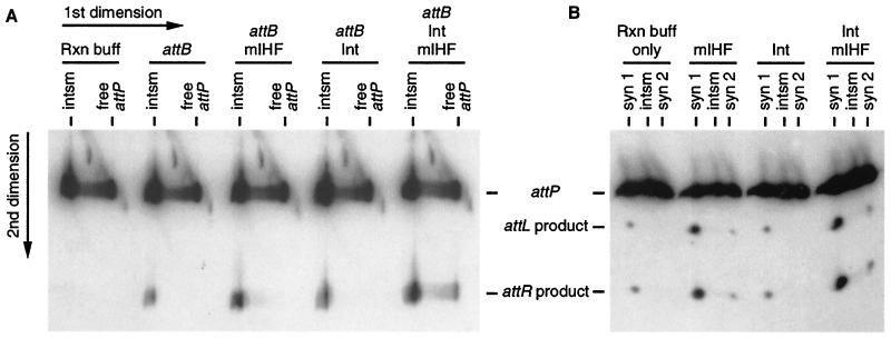
Figure 3.
Activation of recombination in gello. (A) In gello recombination of intasomes. For the first dimension, five reactions similar to the one shown in Fig. 1B lane 3 were run on a native gel to separate intasomes from free attP DNA (labeled at the P1 end), and the lanes were excised. Each gel slice was soaked in reaction (Rxn) buffer either alone or with attB, mIHF, and/or Int-L5 (as indicated). The five gel slices were laid on top of a polyacrylamide gel containing SDS for the second dimension. The last lane (attB/Int/mIHF), in which free attP DNA yields products, demonstrates that attB DNA, mIHF, and Int-L5 do diffuse into the gel slice. (B) In gello recombination of SCs. The intasome, SC1, and SC2 were formed on ice as in Fig. 1B lane 4. The regions of four lanes containing the intasome and both SCs were excised and soaked in reaction buffer (without attB) either alone or with mIHF and/or Int-L5 (as indicated). Recombination products were then identified as in A. For B, complexes were formed by using an attP DNA radiolabeled on both ends, such that both products are visualized. For both A and B, the positions of the intasome and SCs as they ran in the first dimension are labeled on the top, and positions of the DNAs in the second dimension are indicated at the side.

