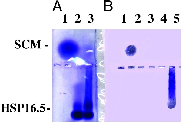
Fig. 4.
Western blot of SCM–Mj HSP16.5 complex. (A) A 0.8% native agarose gel was run as described in Materials and Methods. Lane 1, SCM, 5 μg; lane 2, Mj HSP16.5, 7.4 μg, was heated at 80°C for 20 min and centrifuged for 5 min at 14,000 × g, and the supernatant was loaded; lane 3, SCM–Mj HSP16.5 (1:1 molar ratio) was heated at 80°C for 20 min and centrifuged for 5 min at 14,000 × g, and the supernatant was loaded. (B) A portion of the gel from A was transferred onto nitrocellulose paper and probed with polyclonal antiserum raised against SCM. Lane 1, SCM, 5 μg; lane 3, Mj HSP16.5, 7.4 μg, was heated at 80°C for 20 min and centrifuged for 5 min at 14,000 × g, and the supernatant was loaded; lane 5, SCM–Mj HSP16.5 (1:1 molar ratio) was heated at 80°C for 20 min and centrifuged for 5 min at 14,000 × g, and the supernatant was loaded.

