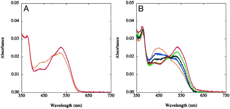
Fig. 2.
Effects of temperature on the UV/visible spectra of methylated wild type (A) and Asp757Glu MetH (649–1227) (B). The MetH fragments (≈3 μM) were diluted into 0.1 M KPi buffer (pH 7.2), at 15°C (red) and 40°C (orange) for the wild-type protein (A), or 15°C (red), 20°C (green), 25°C (black), 30°C (blue), or 40°C (orange) for the Asp757Glu variant (B) and allowed to equilibrate for 2 min before spectral acquisition.

