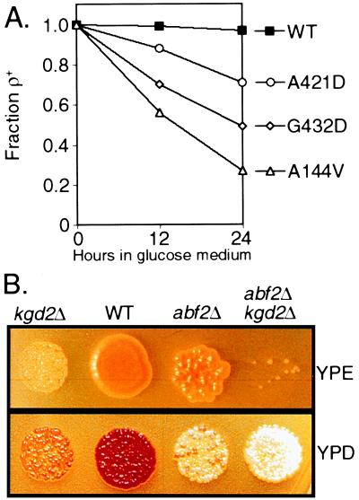
Figure 5.
Genetic analysis of HSP60 and KGD2. (A) Loss of functional ρ+ mtDNA in hsp60-ts mutants. Wild-type and three hsp60-ts mutant strains, listed on the right, were grown overnight in YPGly at 25°C and inoculated into YPD medium at 25°C. The fraction of ρ+ cells in the population present after 12 and 24 h growth was determined in a plating assay by their ability to grow on YPGly plates. (B) Synthetic petite formation between abf2Δ and kgd2Δ mutant strains. Sister spores, prepared as described in Materials and Methods, were plated in equivalent cell numbers on either YPD or YPE medium. Three-fold more cells were plated on YPE than on YPD medium. The loss of ρ+ mtDNA is demonstrated by the decreased number of cells that grow on YPE and the inability of these ade2− strains to develop a red color on YPD.

