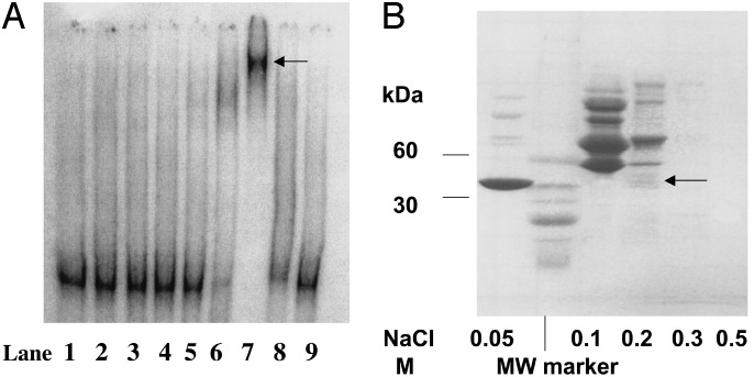
Fig. 1.
(A) Representative EMSA of DNA-binding activity-containing fractions eluted from CM Sepharose. DNA-binding activity-containing fraction eluted from a HiTrap heparin column (0.3 M KCl) was loaded on CM Sepharose (1 mg of protein per ml) and stepwise eluted with buffer A containing NaCl. Lane 1, free 32P-labeled DNA probe; lane 2, effluent of CM Sepharose column; lanes 3 and 4, washes with buffer A; lane 5, 0.05 M NaCl; lane 6, 0.1 M NaCl; lane 7, 0.2 M NaCl; lane 8, 0.3 M NaCl; lane 9, 0.5 M NaCl. (B) Representative Tris/Glycine/SDS/4–20% PAGE of the CM Sepharose fractions that were dialyzed against 10 mM Tris·HCl buffer, pH 6.6 (10 μg of protein per well). The gel was stained with Coomassie blue. The protein bands in the lane designated as 0.2 M NaCl were cut out, and tryptic digests were prepared as described in Materials and Methods.

