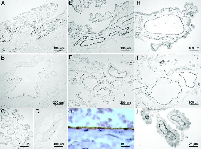
Fig. 4.
P-selectin (A and B) and E-selectin (E and F) immunoreactivity was detected in large venules in choroid plexus (A and E) and SAS (B and F) from individuals who died without neurological or inflammatory disorders. The staining was predominantly detected in large vessels in the choroidal stroma, whereas smaller vessels in choroid plexus villi were negative (C; staining shows P-selectin). In inflammatory parenchymal lesions, punctate P-selectin immunoreactivity was localized to intravascular platelets, whereas endothelial cells were negative (D). The continuous and linear distribution of P-selectin immunoreactivity along the vascular endothelium indicated surface expression of the protein (G; staining shows vessel in choroid plexus). ICAM-1 expression was detected in endothelial cells at multiple localizations in the noninflamed CNS. Shown is characteristic staining of small vessels in the choroid plexus villi (J), large venules in choroidal stroma (H), and SAS (I).

