Figure 1.
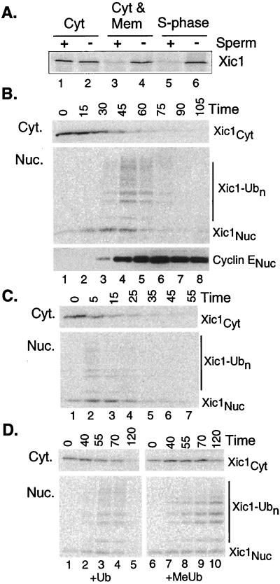
Xic1 is rapidly transported into and ubiquitinated in the nucleus. (A) Destruction of Xic1 requires formation of nuclei. 35S-labeled IVT Xic1 was added to the indicated extract fraction(s) plus or minus sperm DNA. Reactions were processed and analyzed as described in Materials and Methods. Light microscopy confirmed that nuclei formed only in S-phase extract and in the reconstituted cytosolic and membrane fractions (Cyt and Mem). (B) Xic1 is ubiquitinated in the nucleus. Reactions were prepared as in A with sperm DNA, separated into the cytoplasmic and nuclear fractions at the indicated times, and processed as described in Materials and Methods. Samples were analyzed by PhosphorImaging or Western blotting with anti-cyclin E antibody. Subtypes of Xic1 are indicated: Xic1Cyt is the cytoplasmic fraction, Xic1-Ub0 is the unubiquitinated nuclear fraction, and Xic1-Ubn is the ubiquitinated nuclear fraction. The amount of added IVT Xic1 did not measurably affect the normal time course of DNA replication. (C) Xic1 destruction begins rapidly in preformed nuclei. Sperm and energy were mixed with interphase extract and incubated for 50 min to allow nuclei to form. After nuclear formation was confirmed by microscopy, IVT Xic1 and additional extract were added to the reaction (t = 0 min). Samples were removed at indicated times and processed as in B. (D) The modified forms of Xic1 are ubiquitinated. Reactions were prepared as in A and ubiquitin (Ub) or methylated ubiquitin (MeUb) were added and processed at indicated times as in B. To assess the overall effect of MeUb on destruction, comparison of the summed amount of cytoplasmic (Cyt) and nuclear (Nuc) Xic1 remaining was quantitated to be more than 7-fold greater in the sample with added MeUb.

