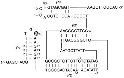
Figure 2.
Secondary structural model of the 10-28 N-glycosylase. The site of depurination (G17) is highlighted. For multiple-turnover experiments, the DNA was divided within the P2 stem after residue A35. Other nucleotide positions that are discussed in the text are numbered. See supplemental Fig. 5 for the sequences of selected variants of the 10-28 clone.

