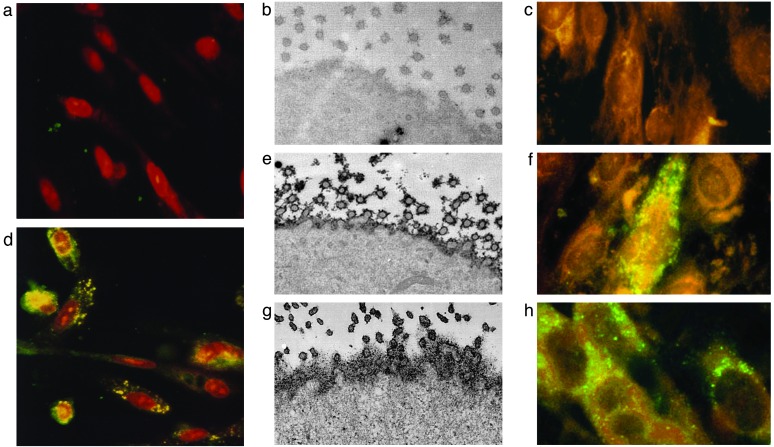
Fig. 3.
Normal BEC develop the phenotypic manifestation of PBC when incubated with PBC patient's lymph node homogenates. (a–c) Before coculture, studies in normal BEC show no AMA staining by immunofluorescence (a), no cell surface expression of PDC-E2 by immunoelectron microscopy (b), and no evidence of viral proteins by immunofluorescence (c). (d–g) After coculture with PBC patient's lymph node homogenates, the BEC develop aberrant PDC-E2 expression after 7 days in culture (d), with cell surface AMA reactivity on BEC (e) that is similar to that seen in PBC BEC (g), and evidence of cytoplasmic localization of betaretrovirus capsid protein (f). (h) Normal BEC incubated with supernatant from MMTV-producing MM5MT cells also show a similar punctate, cytoplasmic signal from the anti-p27CA immunofluorescence.

