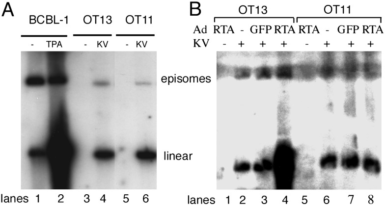
Fig. 3.
Gardella gel analysis of KSHV genomic DNA. (A) OT13 cells (lanes 3 and 4) or OT11 cells (lanes 5 and 6) were mock-infected (lanes 3 and 5) or KSHV-infected (lanes 4 and 6). At 48 h postinfection, infected cells (2 × 10⁶ cells) were subjected to Gardella gel electrophoresis. As a control for linear and circular KSHV DNA, BCBL-1 cells uninduced (-) or induced (+) with phorbol 12-myristate 13-acetate (PMA) were loaded (lanes 1 and 2). The linear DNA observed in latently infected cells (lanes 1, 4, and 6) likely derives primarily from fragmentation of circular episomes during handling. (B) KSHV genomic DNA failed to replicate after RTA induction in KSHV-infected OT11 cells. OT13 (lanes 1–4) or OT11 (lanes 5–8) were mock-infected (lanes 1 and 5) or KSHV-infected (lanes 2–4 and 6–8), followed 2 h later by either Ad-GFP (lanes 3 and 7) or Ad-RTA (lanes 1, 4, 5, and 8) infection. Cells were then subjected to Gardella gel analysis 2 days later.

