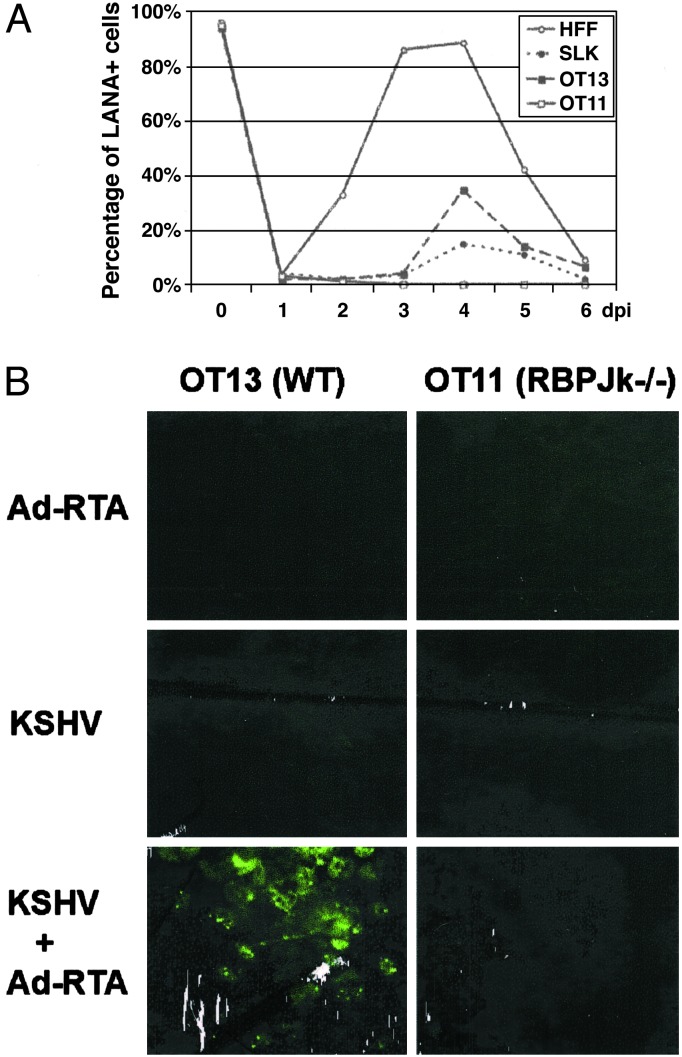
Fig. 5.
Comparison of virus release from induced cells. (A) Time-course analysis of virus release from induced human and mouse cell lines. HFF, SLK, OT13, and OT11 were KSHV-infected for 2 h, followed by Ad-RTA superinfection. Supernatants were collected every day and concentrated by ultracentrifugation. The concentrated virus was then used to infect TIME cells, which were stained for LANA at 48 h postinfection. Serial 2-fold dilutions of the concentrated virus were used in the infectivity assay to ensure that the virus titer is in linear range. The percentage of LANA-positive cells obtained with the undiluted stock from each cell type is plotted against time (days postinfection). ○, HEF; •, SLK; ▪, OT13; □, OT11. (B) No infectious KSHV particles were released after RTA induction in KSHV-infected OT11 cells. OT13 (Left)or OT11 (Right) cells were infected with Ad-RTA alone (Top), KSHV alone (Middle), or KSHV plus Ad-RTA (Bottom). Six days postinfection, supernatants were collected from each culture, concentrated equivalently, and used to infect HFF cells. Two days postinfection, HFFs were stained for LANA expression by immunofluorescence assay.

