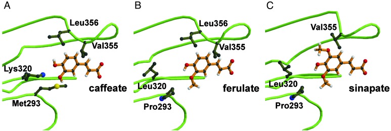
Fig. 2.
3D models of SBPs of different At4CL2 variants. For each enzyme variant the bulkiest substrate that can be fitted into the respective SBP is depicted. In comparison to Fig. 1, the viewpoint has been changed for a better visualization of the respective substrate molecules. In addition, the complexity of the presentation has been reduced by focusing on four amino acid residues of special consideration. (A) SBP of WT enzyme with caffeic acid. (B) SBP of double mutant M293P+K320L with ferulic acid. (C) SBP of triple mutant M293P+K320L+ΔL356 with sinapic acid.

