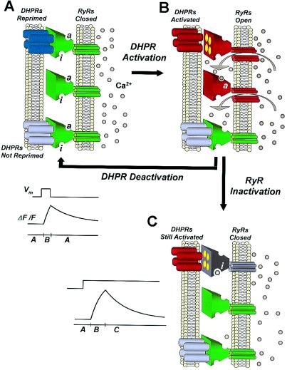
Figure 5.
Model of SR Ca2+ release channel activity underlying sparks terminated by voltage-dependent and voltage-independent mechanisms. Interactions between the ryanodine receptor (RyR) Ca2+ release channel in the SR membrane (right in each panel) and both the voltage sensor (DHPR tetrad) in the t-tubular membrane (left in each panel) and local Ca2+ (gray dots) are shown, with Ca2+ activation (a) and inactivation (i) sites on each RyR indicated in A. In A, all RyRs (green) are closed. The top RyR is coupled to a single reprimed but not activated DHPR tetrad (blue), which is thus available for activation by depolarization. The middle RyR is not coupled to DHPRs but is available for Ca2+ activation secondary to voltage-activated Ca2+ release. The lower RyR is coupled to a nonreprimed DHPR (chartreuse) and thus is unavailable for voltage activation. On depolarization (B), and the resulting DHPR activation (red), the RyR channel coupled to the activated DHPR tetrad opens (red) to conduct Ca2+, which subsequently activates (a) the neighboring non-DHPR coupled channel (red) by Ca2+-induced Ca2+ release. Both open channels may contribute to the generation of a Ca2+ spark. If the depolarization is maintained (C), the DHPR-coupled RyR closes (gray) by a voltage-independent mechanism, probably related to Ca2+ -induced inactivation (i) of the RyR despite the continued activating influence of the voltage sensor (red). If the depolarization is terminated before the RyR is inactivated, the DHPR closes the RyR by voltage sensor deactivation (e.g., B to A; DHPR deactivation), resulting in a Ca2+ spark of briefer rise-time and smaller amplitude. Idealized ΔF/F time courses of a Ca2+ spark generated by DHPR activation with subsequent DHPR deactivation (Upper) or subsequent RyR inactivation (Lower) are shown. The bar beneath each time-course indicates panels (A–C) that represent each phase of the spark ΔF/F time course.

