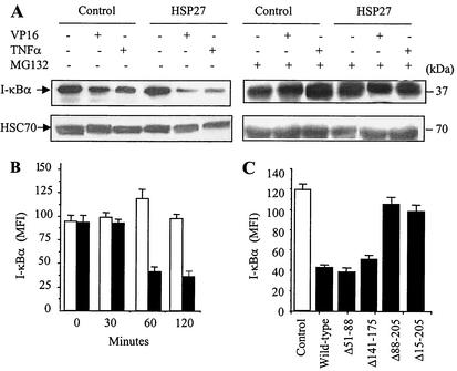
FIG. 6.
HSP27 enhances I-κBα degradation by the proteasome. (A) Control-transfected and HSP27-transfected U937 cells were treated as indicated (VP16, 100 μM, 4 h; ΤNF-α, 20 ng/ml, 4 h; MG132, 25 μM) before monitoring of I-κBα expression by Western blotting. HSC70 served as a loading control. (B) Control-transfected (white bars) and HSP27-transfected (black bars) U937 cells were treated with etoposide (100 μM) for the indicated times before measurement of I-κBα cellular content by flow cytometry (MFI, mean fluorescence index). (C) REG cells were transfected with an empty plasmid (Control) or a plasmid encoding wild-type HSP27 or the indicated deletion mutants of HSP27 and then were treated with etoposide (VP16, 100 μM, 24 h) before measurement of I-κBα cellular content by flow cytometry as described for panel B. Results are the means and standard deviations for four independent experiments.

